Anatomy Of Chest X Ray : Chest Xray Anatomy Labeled Clinical Radiology Anatomy Grepmed / In fact every radiologist and pulmonary physician should be an expert in chest film reading.
Anatomy Of Chest X Ray : Chest Xray Anatomy Labeled Clinical Radiology Anatomy Grepmed / In fact every radiologist and pulmonary physician should be an expert in chest film reading.. In this article we will focus on: Doctors use them to diagnose problems. Each of these anatomical structures should be viewed using a systematic approach. Gillian lieberman forthe harvard 62. Major structures are shown in fig.
A free large database of high quality radiology cases with differential diagnoses and mnemonics to help with board. Each of these anatomical structures should be viewed using a systematic approach. Living anatomy of the chest for 1st year medical students original version compiled by dr. Doctors use them to diagnose problems. A collection of anatomy notes covering the key anatomy concepts that medical students need to learn.
Structure and function of the shoulder complex. Major structures are shown in fig. You have completed this module. Air spaces normally seen in. Therefore, knowing the basics and pathologies in the ed setting is very important.
Free Ai Software For Covid 19 Triage On Chest X Rays Innovation Origins from innovationorigins.com Living anatomy of the chest for 1st year medical students original version compiled by dr. Therefore, knowing the basics and pathologies in the ed setting is very important. In fact every radiologst should be an expert in chest film reading. A free large database of high quality radiology cases with differential diagnoses and mnemonics to help with board. This imaging method can also check how a patient is responding to specific treatments. Doctors use them to diagnose problems. Published 2011 by blackwell publishing ltd. Air spaces normally seen in.
In this article we will focus on:
Many clinical conditions can be evaluated by this simple radiology test. In fact every radiologist and pulmonary physician should be an expert in chest film reading. Both lungs should be well expanded and similar in volume. Major structures are shown in fig. It first appears too complicated to read the chest xrays because we barely know what. These grooves contain the neurovascular bundles that. The interpretation of a chest film requires the understanding of basic principles. A collection of anatomy notes covering the key anatomy concepts that medical students need to learn. Therefore, knowing the basics and pathologies in the ed setting is very important. Common symptoms that can be diagnosed using chest. Doctors use them to diagnose problems. Published 2011 by blackwell publishing ltd. Each of these anatomical structures should be viewed using a systematic approach.
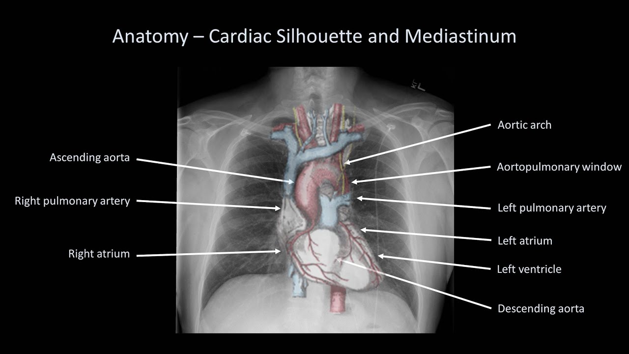
In this article we will focus on: There are also important structures that are obscured or become visible. Heart abnormalities, including fluid around the heart (pericardial effusion), an enlarged heart (cardiomegaly), heart failure, or abnormal anatomy of the heart can be. Labeled chest radiographs teaching radiologic anatomy with a level of detail appropriate for medical students. Living anatomy of the chest for 1st year medical students original version compiled by dr.
Medzcool On Twitter Can You Identify And Name All These Structures On A Chest X Ray Learn More About The Anatomy Of A Chest X Ray Here Https T Co 9ucnjtxkex Radiology Anatomy Medstudent Medicalstudent Usmle Nursing from pbs.twimg.com The chest exam is performed more frequently than any other exam in the imaging department. However, finding problems that are often a/w arrhythmias, such as cardiac enlargement and lung disease, should alter one to the possibility of arrhythmias. Therefore, knowing the basics and pathologies in the ed setting is very important. Is there any inhaled foreign body? Both lungs should be well expanded and similar in volume. Structure and function of the shoulder complex. Living anatomy of the chest for 1st year medical students original version compiled by dr. Each of these anatomical structures should be viewed using a systematic approach.
Each of these anatomical structures should be viewed using a systematic approach.
In this article we will focus on: It is almost always the first imaging study ordered to evaluate for pathologies of the thorax, although further diagnostic imaging, laboratory tests. Conclusion of living anatomy of the chest congratulations! Air spaces normally seen in. Chest radiographs are the most common film taken in medicine. Structure and function of the shoulder complex. You have completed this module. Major structures are shown in fig. It first appears too complicated to read the chest xrays because we barely know what. Is there any inhaled foreign body? Published 2011 by blackwell publishing ltd. The chest exam is performed more frequently than any other exam in the imaging department. Evaluation of a chest radiograph may appear to be simple, but is in fact a complex task requiring careful observation, sound understanding of chest anatomy, and knowledge of the principles of physiology and pathology.
Major structures are shown in fig anatomy of chest. You have completed this module.


0 Komentar